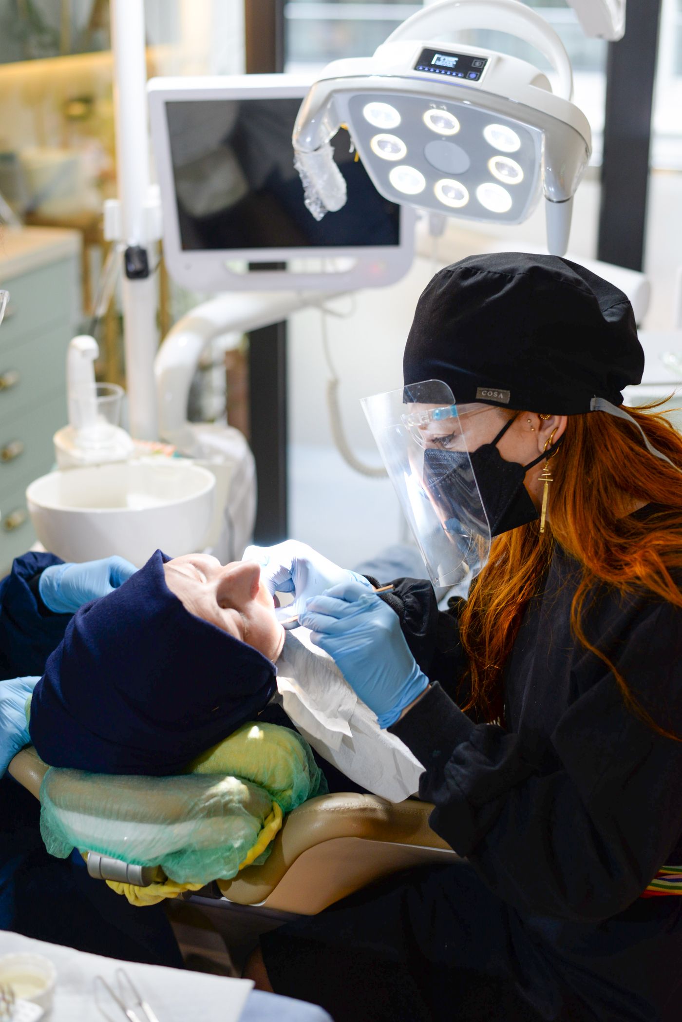
Dentacare describes digital impressions as a modern alternative to conventional impression material. A small scanning camera records the teeth and gums and software converts the images into a detailed three-dimensional model.
Why digital impressions are used
- A cleaner, more comfortable scanning experience
- Detailed digital models for planning and fabrication
- Immediate on-screen review
- Efficient transfer to the dental laboratory or design workflow
- Potentially fewer adjustments through improved accuracy
How the scan works
A wand-like intraoral scanner is moved around the teeth while it captures many images. Software joins the information into a 3D model that can support the design of crowns, bridges, veneers, night guards or orthodontic appliances.
CAD/CAM restorations
Computer-aided design and manufacturing can use the digital model to plan a restoration that fits the patient’s tooth and bite. The restoration may then be milled from materials such as ceramic or zirconia, depending on the clinical requirement.
From scanning to a 3D model
An intraoral scanner captures a rapid series of images as the tip moves around the teeth. Software identifies overlapping details and assembles them into a three-dimensional surface model. The operator can rotate the model, magnify areas and rescan information that is missing.
Unlike a conventional impression, the digital model can be reviewed immediately. This does not remove the need for careful technique: moisture control, visibility of the preparation and accurate capture of the bite remain essential.
Where digital impressions are useful
Digital scans can support crowns, bridges, veneers, implant restorations, retainers, aligners, night guards and diagnostic records. Files can be transferred to the laboratory without physically transporting an impression and can be stored for comparison where the system permits.
Not every clinical situation is best captured digitally. Deep areas below the gum, extensive mobile tissue and some full-denture cases may still require conventional or combined techniques.
- Immediate visual quality check
- No tray filled with impression material
- Efficient communication with the laboratory
- Useful digital records for planning
- Integration with computer-aided design
- Potentially easier patient explanation
What CAD/CAM adds
CAD software helps the dentist or technician design the restoration on the digital model. CAM equipment then manufactures it by milling or printing an appropriate material. The restoration still needs clinical checks for fit, contact, bite, colour and cleanability.
Technology improves the workflow but does not make decisions by itself. Diagnosis, material selection, tooth preparation and final adjustment continue to depend on trained clinical judgement.
Frequently Asked Questions
Does scanning expose me to radiation?
An optical intraoral scan uses light to record surface shape and is different from an X-ray. Separate radiographs may still be needed for information beneath the surface.
Is a digital scan uncomfortable?
Most patients find the small scanning tip easier than impression material, although the mouth must remain open while all areas are recorded.
Can a crown be made on the same day?
Some clinics and cases support a same-day workflow, while others require laboratory fabrication. Suitability depends on equipment, material and clinical complexity.